
Walk onto any ward in the country and you’ll find oxygen running — often without a prescription, often at the wrong flow rate, often in a patient who doesn’t need it. As a student nurse, you’ll be asked to “put some oxygen on” more times than you can count. This guide explains why that phrase should make you pause, what the correct target saturations are, which device to use, and the one patient group where giving too much oxygen can be genuinely dangerous.
Oxygen is so ubiquitous — piped into every wall, available at every bedside — that it’s easy to treat it as harmless, like turning up a light. It isn’t. Oxygen is a drug. It has indications, contraindications, side effects, and a correct dose. It must be prescribed. It must be monitored. And it must be weaned off when no longer needed. The British Thoracic Society estimates that up to a third of hospital patients receive oxygen at some point — and audits consistently show that around half of those prescriptions are wrong: no target saturation, no device specified, or simply no prescription at all.
For a student nurse, oxygen is one of the highest-yield skills to master. It comes up in every placement, every OSCE, every interview, and every deteriorating patient scenario. And the rules are simple — once you know them. Let’s break it down.
Part 1: What You’re Actually Treating — Hypoxaemia vs Hypoxia
Before you reach for the mask, know the difference between two words that get used interchangeably but mean very different things:
- Hypoxaemia = low oxygen in the blood — measured by pulse oximetry (SpO₂) or arterial blood gas (PaO₂). This is what oxygen treats.
- Hypoxia = low oxygen at the tissues — the organs aren’t getting enough. This can happen with a normal SpO₂ (e.g. severe anaemia, carbon monoxide poisoning, shock with poor perfusion). Oxygen alone won’t fix it.
The key point: You give oxygen to correct hypoxaemia — to get the saturations into the target range. If the saturations are already in range, more oxygen does not help and may harm. A patient who is “breathing fast” but has normal sats does not need oxygen — they need assessment for the cause (pain, anxiety, sepsis, acidosis). This is where working through ABCDE properly saves you from a reflex “oxygen on” response.
Part 2: Target Saturations — The Two Ranges You Must Know
The single most important fact in oxygen therapy: there is not one target. There are two, and picking the wrong one for the wrong patient is the classic, examinable, potentially fatal mistake.
| Patient Group | Target SpO₂ | Examples |
|---|---|---|
| Most acutely unwell adults | 94–98% | Sepsis, pneumonia, asthma, heart failure, post-op, trauma — the default for everyone not in the at-risk group |
| At risk of hypercapnic (type 2) respiratory failure | 88–92% | COPD, obesity hypoventilation syndrome, neuromuscular disease (e.g. MND, muscular dystrophy), chest wall deformity (e.g. kyphoscoliosis), cystic fibrosis |
| Carbon monoxide poisoning, cardiac arrest, resuscitation | 100% (until definitive treatment) | Resuscitation situations — high-flow non-rebreather, per Resuscitation Council UK |
⚠️ The Critical Distinction
A patient with known COPD who is acutely unwell does not automatically get 88–92% — if they’re critically ill and peri-arrest, you give high-flow oxygen and sort the target out later. But for the stable or moderately unwell COPD patient, the target is 88–92%. When in doubt: start at 88–92% in any patient with known COPD or a “smoker’s chest,” and confirm with an ABG within 30–60 minutes. Never leave a COPD patient on uncontrolled high-flow oxygen without monitoring.
Note also: in recent years, evidence has shifted against routine high-flow oxygen in conditions where it was once standard. We now know that giving high-flow oxygen to patients with acute stroke or myocardial infarction who are not hypoxic can worsen outcomes — oxygen causes vasoconstriction and increases oxidative stress. So the rule is now universal: oxygen only if hypoxic, titrated to target. This is exactly why our stroke recognition guide specifies oxygen only if saturations fall below 94%.
Part 3: Oxygen Delivery Devices — Which One, Which Flow
There are more devices than you think, and they deliver very different concentrations of oxygen (FiO₂ — fraction of inspired oxygen). Pick the wrong one and you’re either under-treating a hypoxic patient or over-oxygenating an at-risk one.
| Device | Flow Rate | Approx. FiO₂ | When To Use |
|---|---|---|---|
| Nasal Cannulae | 1–6 L/min | ~24–44% (roughly +4% per L/min above room air) | Low-requirement patients, long-term therapy, weaning. Comfortable, allows eating/talking. Above 6 L/min it dries the nose and delivers little extra benefit |
| Simple Face Mask (Hudson) | 5–10 L/min (never below 5) | ~40–60% | Moderate requirements. Below 5 L/min the patient re-breathes exhaled CO₂ — never run a simple mask at 2–4 L/min |
| Venturi (Fixed-Performance) Mask | Set per valve (e.g. 2, 4, 8, 15 L/min) | Fixed: 24, 28, 35, 40, 60% | Controlled oxygen — essential for COPD/at-risk patients. Each colour-coded valve delivers a precise FiO₂ at a specified flow rate; the flow must match the valve or the FiO₂ is wrong |
| Non-Rebreather (Reservoir) Mask | 10–15 L/min | ~60–85%+ | Emergency, critical illness, trauma, shock. Inflate the reservoir bag before applying. A deflated reservoir delivers no benefit. Short-term only — requires monitoring |
| Tracheostomy Mask / HME | Via tracheostomy | Variable | Tracheostomy patients — humidification is essential (HME or heated). Never put a standard face mask over a tracheostomy |
| High-Flow Nasal Oxygen (HFNO — e.g. Optiflow) | Up to 60–70 L/min, heated & humidified | Precise, up to ~100% | Type 1 respiratory failure, post-extubation, specialist wards/ED/ITU. Delivers high flow with precise FiO₂ and PEEP-like effect — specialist initiation |
| CPAP / BiPAP (Non-Invasive Ventilation) | Specialist set-up | Controlled | CPAP: cardiogenic pulmonary oedema, type 1 failure. BiPAP: hypercapnic type 2 failure (COPD exacerbation). Only initiated by specialists — your role is monitoring and escalation |
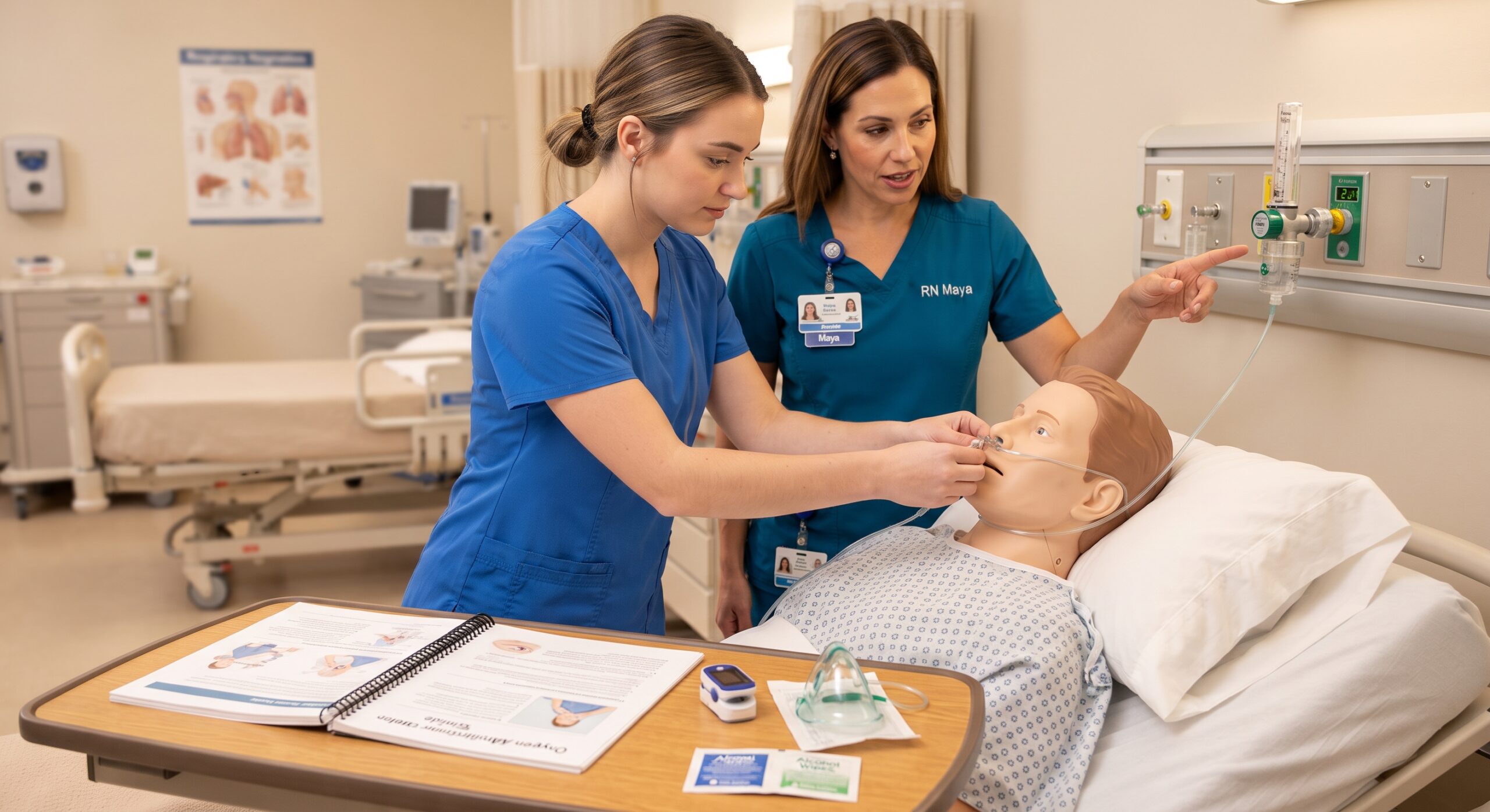
Oxygen delivery equipment. Whatever device you choose, it must be prescribed with a target saturation range, flow rate, and regular monitoring plan — oxygen is a drug, not a bedside comfort measure.
Quick device rules students always forget:
- Nasal cannulae max 6 L/min — beyond that, you’re just drying the nasal passages with no real FiO₂ gain
- Simple face mask minimum 5 L/min — below that, exhaled CO₂ builds up in the mask
- Non-rebreather: inflate the bag first — always. A flat reservoir is a useless mask
- Venturi: flow rate must match the valve — the percentage on the valve is only accurate at the stated flow. Check the label on the valve, every time
- Humidify for high flow or prolonged use — see Part 5
Part 4: The COPD Danger — Why Too Much Oxygen Kills
This is the one that gets examined, and the one that genuinely harms patients. In patients with chronic CO₂ retention (classically severe COPD), giving high-concentration oxygen can push them into type 2 respiratory failure — drowsiness, rising CO₂, acidosis, and potentially coma or death. Two mechanisms are at work:
- V/Q mismatch: Hypoxic pulmonary vasoconstriction normally redirects blood to well-ventilated areas of the lung. High FiO₂ abolishes this, sending blood to poorly ventilated areas and worsening CO₂ elimination.
- The Haldane effect: Oxygenated blood carries CO₂ less well, so high FiO₂ increases the CO₂ content of blood directly.
(The old “hypoxic drive” explanation — that oxygen removes the stimulus to breathe — is a minor contributor, not the main one. But the clinical takeaway is identical: high oxygen in at-risk patients causes CO₂ retention.)
⚠️ What CO₂ Retention Looks Like
A COPD patient who was breathless but alert, put on high-flow oxygen, becomes drowsy, confused, flushed, with a bounding pulse and a slow respiratory rate. Their saturations may look “great” (99–100%) — that’s the trap. High sats + drowsy = think CO₂ retention. Reduce the oxygen, sit them up, call for senior help immediately, and prepare for BiPAP/NIV. An ABG will confirm a rising PaCO₂ and falling pH.
The safe approach for any patient with known COPD or a heavy smoking history: start with a Venturi mask at 28% (or 24% if very high risk), target 88–92%, and arrange an ABG within 30–60 minutes. If they’re critically unwell and peri-arrest, give high-flow oxygen — but tell the team the moment they arrive so the target can be corrected. Never leave them on it unattended.
Part 5: Humidification — When Dry Oxygen Becomes a Problem
Oxygen from the wall or a cylinder is dry. Your upper airway normally humidifies and warms inspired air to 100% humidity at body temperature. Bypass that (via a mask at high flow, or a tracheostomy) and you deliver cold, dry gas straight into the lower airway. The result: cilia stop working, secretions thicken and crust, mucous plugs form, atelectasis develops, and the airway becomes irritated.
Humidify when:
- Oxygen flow is high (typically >4 L/min via mask, or any high-flow system)
- Therapy is prolonged (more than a few hours, or days)
- Secretions are thick or the patient is struggling to clear them
- The patient has a tracheostomy or artificial airway — humidification is mandatory (HME — heat and moisture exchanger — or heated humidifier)
Options: simple bubble humidifier bottles for low-flow mask use; heated humidifier circuits for high flow and ventilators; HME “noses” for tracheostomies. For tracheostomy patients, an HME should be in place at all times when not on active humidification — a bare tracheostomy open to dry air is a recipe for mucous plugging and distress.
Part 6: Nebulisers — Air-Driven vs Oxygen-Driven
Bronchodilator nebulisers (salbutamol, ipratropium) are given via a nebuliser driven by either air or oxygen. The choice matters:
- Asthma / no CO₂ risk: Oxygen-driven nebulisers are fine — target 94–98%. In a severe asthma attack, oxygen-driven nebulisers are standard.
- COPD / at risk of hypercapnia: Use air-driven nebulisers where possible. If oxygen is needed concurrently, give it via nasal cannulae alongside (controlled) rather than driving the nebuliser with high-flow oxygen. Monitor sats and consciousness closely.
The placement reality: Many wards only have oxygen outlets at the bedside, so air-driven nebulisers require a compressor pump. If the only option is oxygen-driven in a COPD patient, give it — but monitor the patient closely, don’t leave them, and flag to the team that ABG monitoring is needed. Don’t withhold a needed bronchodilator over the drive gas; just be aware of the risk.
Part 7: Prescribing Oxygen — What a Valid Prescription Looks Like
Because oxygen is a drug, it must be prescribed on the drug chart or the designated oxygen prescription section. A valid oxygen prescription includes all four of these — anything less is incomplete:
The 4 Essentials of an Oxygen Prescription
- Target saturation range — e.g. “94–98%” or “88–92% (COPD).” This is the single most important bit. Without a target, nobody knows when to turn it up or down.
- Device — nasal cannulae, Venturi 28%, simple mask, non-rebreather, etc.
- Flow rate / FiO₂ — e.g. “2 L/min via nasal cannulae” or “Venturi 28% at 4 L/min.”
- Monitoring & weaning plan — how often to check sats, when to reduce, and to wean to room air as soon as possible.
If you find oxygen running without a prescription — and you will — it is not your job to prescribe it, but it is your job to flag it to a doctor or prescriber and document that you did so. “Put some oxygen on” is a verbal instruction, not a prescription. Always confirm the target. This is medicines management, and it’s covered by the NMC Code — our defensive documentation guide covers exactly how to record these conversations.
Part 8: Starting, Monitoring & Weaning — Step by Step
Oxygen is not a “set and forget” treatment. It is titrated — started, checked, adjusted, and weaned. Here’s the cycle:
- Assess. ABCDE. Check sats, respiratory rate, work of breathing, colour, consciousness. Full observations including NEWS2. If sats below target, escalate and start oxygen. If the patient is severely unwell, this is part of the emergency response — see our Panic Zone for the emergency pathway.
- Prescribe. Ensure a valid prescription (target, device, flow, monitoring plan) exists or is being written.
- Apply the correct device. Inflate reservoirs if non-rebreather. Set flow to match the device/valve.
- Re-check within 5 minutes. Are sats rising into target? If not, increase stepwise (under instruction) — don’t just leave it.
- ABG if indicated. If at-risk of hypercapnia, if not improving, or if critically unwell — ABG within 30–60 minutes. Repeat after any change in at-risk patients.
- Monitor regularly. Continuous pulse oximetry for the unstable; regular spot checks for the stable. Document sats, device, and flow on the observation chart.
- Wean early. As soon as sats are stable in range on a low flow, reduce — drop a Venturi step, or reduce nasal cannulae by 1 L/min. Re-check in 5–10 minutes. Aim for room air. Oxygen left on at 2 L/min “because it’s always been on” is one of the commonest sources of avoidable harm.
The weaning rule of thumb: If a patient is sat comfortably in chair, eating, chatting, with sats 96% on 2 L/min nasal cannulae — they almost certainly don’t need it anymore. Wean. The longer unnecessary oxygen runs, the more it dries secretions, suppresses appetite (nasal cannulae get in the way of eating), and delays discharge. Weaning is a nursing skill — own it.
Part 9: Pulse Oximetry — What It Does (and Doesn’t) Tell You
The pulse oximeter is your main monitoring tool — but it has important limitations. A “normal” reading can be falsely reassuring, and a low reading can be artefactual. Know both:
| Limitation | What Happens | What To Do |
|---|---|---|
| Poor perfusion / shock / hypothermia | No trace, or reading drops out — the probe can’t detect pulsatile flow | Warm the hand, try earlobe/forehead probe, don’t assume the patient is desaturating — but assess them |
| Nail varnish / artificial nails | Inaccurate or unobtainable reading | Remove varnish or use a different finger/earlobe |
| Carbon monoxide poisoning | Carboxyhaemoglobin reads as oxyhaemoglobin — SpO₂ shows ~100% despite severe hypoxia | ABG with CO-oximetry. High-flow 100% oxygen. Suspect in fires, enclosed spaces, headache + confusion |
| Dark skin pigmentation | Pulse oximeters can overestimate saturation in patients with darker skin (FDA warning 2022) — occult hypoxaemia missed | Trust the patient, not just the number. Lower threshold for ABG if clinically unwell. Be aware of the bias |
| Severe anaemia | SpO₂ measures saturation of the haemoglobin present, not oxygen content — can be normal with dangerously low oxygen carriage | Check FBC. A pale, breathless patient with “normal sats” may still be tissue-hypoxic |
| Motion / shivering | Errant readings, dips that aren’t real | Immobilise the probe site, re-check, correlate with clinical state |
The golden rule: treat the patient, not the monitor. A reading that doesn’t match the clinical picture needs re-checking and, if in doubt, an ABG. A patient who looks terrible with a “normal” sats is still terrible. Conversely, a low reading with a good trace and a well-looking patient may be probe error — but verify before dismissing.
Part 10: The Commonest Mistakes — And How to Avoid Them
| Mistake | Why It’s Wrong | The Fix |
|---|---|---|
| No prescription / no target saturation | Oxygen is a drug — unprescribed, unmonitored | Always confirm target 94–98 or 88–92; flag missing prescription |
| High-flow oxygen to a COPD patient | CO₂ retention, type 2 failure, coma | Venturi, target 88–92, ABG within 30–60 min |
| Simple mask at <5 L/min | Re-breathing exhaled CO₂ | Minimum 5 L/min on a simple mask — or switch to cannulae for low flow |
| Nasal cannulae at >6 L/min | No extra FiO₂, just drying and discomfort | Switch to a mask if more oxygen needed |
| Non-rebreather with deflated reservoir | Delivers nowhere near the claimed high FiO₂ | Inflate the bag before applying; check it stays inflated on inspiration |
| Venturi at wrong flow rate | The stated FiO₂ is only accurate at the marked flow | Read the valve label every single time; set flow accordingly |
| No humidification on high/prolonged therapy | Thick secretions, mucous plugs, atelectasis | Humidify; mandatory for tracheostomy |
| Never weaning — “it’s always been on” | Prolonged unnecessary oxygen, delayed discharge, harm | Wean early and often; aim for room air |
| Trusting the sats blindly | Missed occult hypoxaemia, CO poisoning, anaemia, bias | Treat the patient; ABG when in doubt |
| Oxygen for breathlessness with normal sats | Oxygen doesn’t treat the cause; may harm | Assess for cause — pain, anxiety, sepsis, acidosis; oxygen only if hypoxic |
Part 11: Escalation — The SBAR Script for Desaturations
When a patient’s sats drop and they’re not responding to initial oxygen, escalate. Use SBAR — it’s the structure that gets you heard. Here’s the exact script:
S — Situation: “This is [Name], student nurse on [Ward]. I’m with Mr/Mrs [Surname] and their saturations have dropped to [X]% on [current oxygen]. They’re [breathless/drowsy/chest pain].”
B — Background: “Admitted with [reason]. History of [COPD/asthma/heart failure]. Baseline sats [X]%. Oxygen currently [device] at [flow], target [range].”
A — Assessment: “ABCDE: airway patent, respiratory rate [X], using accessory muscles, oxygen increased to [device/flow] per [instruction], sats now [X]%, pulse [X], BP [X], GCS [X], temp [X], NEWS2 [score]. ABG [taken/sent/pending].”
R — Recommendation: “I need you to review them immediately. I think they need senior review / ITU outreach / may need NIV. Can you come now?”
Remember that oxygen is part of the immediate response to sepsis — the Sepsis Six includes high-flow oxygen to maintain SpO₂ ≥94% as one of the six immediate actions. If sepsis is on your differential, oxygen is one of the first things you do — alongside cultures, fluids, lactate, and antibiotics.
Part 12: Documentation — Record Everything
Every oxygen intervention must be documented: when it was started, device, flow rate, target range, who prescribed it, sats before and after, ABG results, weaning steps, and any concerns raised. If a patient deteriorates on oxygen and there’s no record of the target or the monitoring, it looks like negligence — even if the care was good. Our defensive documentation guide covers this in full, and the same principles apply to oxygen as to any other drug.
Final Thought
Oxygen is the gateway drug of hospital care — so normal, so available, that it stops feeling like a drug at all. That’s exactly when it becomes dangerous. The best nurses I’ve worked with treat oxygen with the same respect as any other prescription: they check the target, pick the right device, monitor the response, and wean it off as soon as they can.
You will be asked to “just put some oxygen on” hundreds of times as a student. Each time, pause for two seconds and ask: what’s the target? which device? who prescribed it? Those two seconds make you a safer nurse. And one day — possibly on a COPD patient who would have drifted into CO₂ narcosis — those two seconds will be the difference between a routine shift and a bad one.
Oxygen saves lives. Used wrongly, it ends them. Know the difference.
📚 References & Guidance
- British Thoracic Society (2017) BTS Guideline for Oxygen Use in Adults in Healthcare and Emergency Settings. O’Driscoll BR et al. — the definitive UK guideline; target saturations, devices, prescribing, weaning.
- O’Driscoll BR et al. (2008, updated 2017) “BTS guideline for emergency oxygen use in adult patients.” Thorax / BMJ Open Respiratory Research — evidence base for 94–98 and 88–92 targets.
- Resuscitation Council UK (2021) Quality Standards for Resuscitation — Oxygen in Acute Illness & Cardiac Arrest. High-flow 100% oxygen during resuscitation.
- NICE (2019, updated) NG114: Chronic Obstructive Pulmonary Disease — Management. Oxygen targets and nebuliser guidance in COPD.
- NICE (2016) NG5: Sepsis — Recognition, Diagnosis & Early Management. Sepsis Six includes oxygen to maintain SpO₂ ≥94%.
- National Patient Safety Agency / NHS Improvement (2009 onwards) Oxygen Safety Alerts — Tubing Misconnections & Prescribing. Patient safety risks of oxygen delivery systems.
- Medicines and Healthcare products Regulatory Agency (MHRA) Medical Device Alerts — Pulse Oximeters. Accuracy limitations.
- US Food & Drug Administration (2022) FDA Safety Communication: Pulse Oximeter Accuracy Limitations, Including Skin Pigmentation. Occult hypoxaemia risk in darker skin.
- NMC (2018) The Code: Professional Standards. Medicines management — oxygen as a prescribed drug; Paragraphs 3.1–3.14.
- WHO (2017) Patient Safety Solutions — Oxygen Therapy. Global safety guidance.
- Avoid trials / DETO2X-AMI (2014–2017) Oxygen Therapy in Suspected Acute Myocardial Infarction & Stroke. Evidence that routine high-flow oxygen in non-hypoxic patients may worsen outcomes.
- National Confidential Enquiry into Patient Outcome and Death (NCEPOD) Respiratory failure audits — oxygen prescribing failures in ward settings. Consistent finding: ~50% of oxygen prescriptions incomplete.
Written by: Daniel Hancock, Registered Nurse
Focus: Oxygen as a drug, target saturations, delivery devices, COPD/hypercapnia risk, humidification, nebulisers, prescribing, weaning, pulse oximetry limitations
Published: October 2026 | Last reviewed: October 2026
Intended for: UK student nurses & newly qualified nurses — ward-based oxygen therapy
Always follow your trust’s specific oxygen policy and the BTS guideline. Target saturations, device choice, and weaning must be individualised — especially in COPD, neuromuscular disease, and other hypercapnia-risk groups. This guide is educational and does not replace clinical judgement, senior review, or a valid prescription. If a patient is deteriorating despite oxygen, escalate immediately. Never leave a non-rebreather mask applied with a deflated reservoir, and never run a simple face mask below 5 L/min.
Reinforce Your Acute Care Skills
Oxygen sits at the heart of every deteriorating patient. Pair it with the frameworks and practice that keep you safe under pressure.