
A stroke is the one emergency where your observation skills — the student nurse at the bedside — directly determine whether someone walks out of hospital or spends the rest of their life in a wheelchair. The doctors can’t treat what they haven’t been told about. The scans can’t happen until you press the buzzer. This guide gives you everything you need to recognise a stroke fast, escalate it correctly, and understand what happens in that golden first hour.
Stroke is the fourth leading cause of death in the UK and the single biggest cause of adult disability. Around 100,000 people have a stroke every year — that’s one every five minutes. You will see strokes on placement. You will almost certainly see one missed. And the difference between a good outcome and a devastating one is measured in minutes, not hours.
The phrase you’ll hear over and over is “time is brain.” It sounds like a slogan. It’s not. It’s a physiological fact. Every minute that a large blood vessel in the brain remains blocked, approximately 1.9 million neurons die. That’s 120 million an hour. The longer you wait, the more brain is permanently lost — and the less likely any treatment is to work. So when you suspect a stroke, you do not finish your drug round. You do not wait for the staff nurse to come back from break. You escalate — now.
Part 1: What Actually Is a Stroke?
A stroke (properly called a cerebrovascular accident, or CVA) happens when the blood supply to part of the brain is suddenly interrupted. Brain cells are incredibly greedy — they need a constant supply of oxygen and glucose, and they start dying within minutes without it. There are two fundamentally different types, and the treatment is almost the exact opposite — which is why you can never, ever assume which one it is without a scan.
| Type | What Happens | Proportion | Treatment Direction |
|---|---|---|---|
| Ischaemic Stroke | A clot blocks a blood vessel — either forming in the brain (thrombosis) or travelling from elsewhere (embolism, often from the heart in atrial fibrillation) | ~85% | Clot-busting drugs (thrombolysis) or clot retrieval (thrombectomy) |
| Haemorrhagic Stroke | A blood vessel bursts and bleeds into or around the brain — intracerebral haemorrhage or subarachnoid haemorrhage | ~15% | Stop the bleeding, control blood pressure, sometimes neurosurgery — thrombolysis would kill these patients |
| TIA (Mini-Stroke) | Temporary blockage — symptoms fully resolve, usually within an hour, always within 24 hours | Common | Not a “mini” anything — ~10% go on to have a full stroke within 7 days. Urgent specialist review |
The critical point: Ischaemic and haemorrhagic strokes look identical at the bedside. You cannot tell them apart from the symptoms. Giving thrombolysis (a clot-buster) to someone having a bleed would be catastrophic. That’s why a CT scan is the absolute first priority — and why everything you do before the scan is about stabilisation and speed, not treatment.
What about TIAs?
A Transient Ischaemic Attack is when stroke-like symptoms resolve completely — by definition within 24 hours, though most resolve within an hour. Students (and unfortunately some qualified staff) sometimes dismiss these as “just a TIA.” That is dangerous. A TIA is a warning shot. The risk of a full stroke in the 7 days following a TIA is around 10% — and half of those happen in the first 48 hours. A patient with new TIA symptoms needs urgent specialist assessment, often the same day, and treatment (antiplatelets, statin, blood pressure control) started immediately. If someone has stroke symptoms that resolve, they still need to be seen — urgently. Do not let them go home.
Part 2: FAST & BE-FAST — The Recognition Tools
FAST is the public-facing stroke recognition tool, and it’s the one every member of the multidisciplinary team should know cold. It was developed from the Cincinnati Prehospital Stroke Scale and has been validated repeatedly. Here’s what each letter means — and how to test it properly, not just glance at the patient.
| Letter | Test | What An Abnormality Looks Like |
|---|---|---|
| F — Face | Ask the person to smile or show their teeth | One side droops. Key distinction: a stroke causes forehead-sparing weakness (they can still wrinkle both sides of their forehead). Bell’s palsy affects the whole side, including the forehead |
| A — Arms | Ask them to hold both arms out straight, palms up, eyes closed, for 10 seconds | One arm drifts down or cannot be lifted at all. Check grip strength too — squeeze your fingers with both hands |
| S — Speech | Ask them to say a simple sentence: “You can’t teach an old dog new tricks” or “The sky is blue in Chesterfield” | Slurred speech (dysarthria), wrong words (dysphasia/aphasia), or inability to understand you. Don’t accept “they’re just a bit confused” |
| T — Time | Note the exact time symptoms started — or when they were last seen well — and call 999 / put out the emergency call immediately | This is the single most important piece of information you will record. It determines whether thrombolysis is possible |
Why BE-FAST exists — the strokes FAST misses
Here’s the problem students rarely get taught: FAST was designed for the public to spot common strokes affecting the carotid (anterior) circulation. But around 20% of strokes affect the posterior circulation — the back of the brain, the cerebellum and brainstem — and these often present with no facial droop, no arm weakness, and no speech problem. They present with dizziness, double vision, imbalance, and difficulty swallowing. FAST misses them. That’s why many trusts and ambulance services now use BE-FAST, adding two letters:
- B — Balance: Sudden loss of balance, inability to walk straight, vertigo, or falling to one side. A patient who “just feels dizzy” and can’t walk in a straight line is a stroke until proven otherwise
- E — Eyes: Sudden double vision (diplopia), loss of vision in one eye (amaurosis fugax — a “curtain coming down”), or gaze deviation where the eyes look toward one side and won’t cross the midline
⚠️ The Danger Pattern
An elderly patient who says “I just feel a bit dizzy and sick,” who can’t stand, and whose observations are normal — this is the classic missed posterior circulation stroke. They get diagnosed with a UTI or “just a bit of vertigo” and sit on the ward for 12 hours. If you see this, escalate. Dizziness + new neurological deficit (even subtle) = stroke until proven otherwise.
There are other recognised scales too — the ROSIER scale (Recognition Of Stroke In the Emergency Room) is used in many EDs and includes loss of consciousness/seizure and leg weakness as additional factors. But for the ward nurse, BE-FAST is the tool that will catch the most strokes with the least faff. Learn it, and use it every time you have even a flicker of suspicion.
Part 3: The Hyperacute Response — What Happens & Why Speed Is Everything
When a stroke is suspected, the patient enters the hyperacute stroke pathway. In England, most hospitals are part of a network with a designated Hyperacute Stroke Unit (HASU) — sometimes on your site, sometimes at a neighbouring hospital the patient is blue-lighted to. The pathway runs on strict time targets, and every member of the team has a role. Here’s the timeline you need to understand:
Symptoms begin, or you spot them at the bedside. You note the time of onset (or last seen well), do a quick BE-FAST screen, and call for help. The clock starts now.
ABCDE assessment, check capillary blood glucose (hypoglycaemia mimics stroke), full set of observations including NEWS2, oxygen only if hypoxic, IV access, keep nil by mouth until swallow screened. Call the senior nurse and doctor — or put out the 2222 emergency call if the patient is deteriorating.
NICE guidance (NG128) requires urgent brain imaging — a CT scan within 1 hour of arrival (or immediately if on your ward). CT is fast and excellent at showing blood (haemorrhage), which is the critical first question. It may not show an early ischaemic stroke at all — that’s normal. The point is to exclude a bleed.
If the CT shows no blood, a CT angiogram (CT with contrast looking at the arteries) identifies whether there’s a large vessel occlusion — the clots that cause the worst strokes and are eligible for thrombectomy.
If it’s an ischaemic stroke, no bleed, and the patient is within 4.5 hours of onset (with no contraindications), they may be offered intravenous thrombolysis — usually alteplase, increasingly tenecteplase — which dissolves the clot. The earlier it’s given, the better the outcome. Every 15 minutes of delay meaningfully reduces the chance of a good recovery.
For large vessel occlusions, mechanical thrombectomy — a specialist interventional radiologist threads a device through the arteries and physically removes the clot — can be offered up to 24 hours after onset in selected patients, if imaging shows salvageable brain tissue (the “penumbra”). This is the single biggest advance in stroke care in a decade.
The patient goes to a specialist HASU for close monitoring: blood pressure control, swallow screening, antiplatelets/statins, early mobilisation, and complication prevention. The first 72 hours are critical.
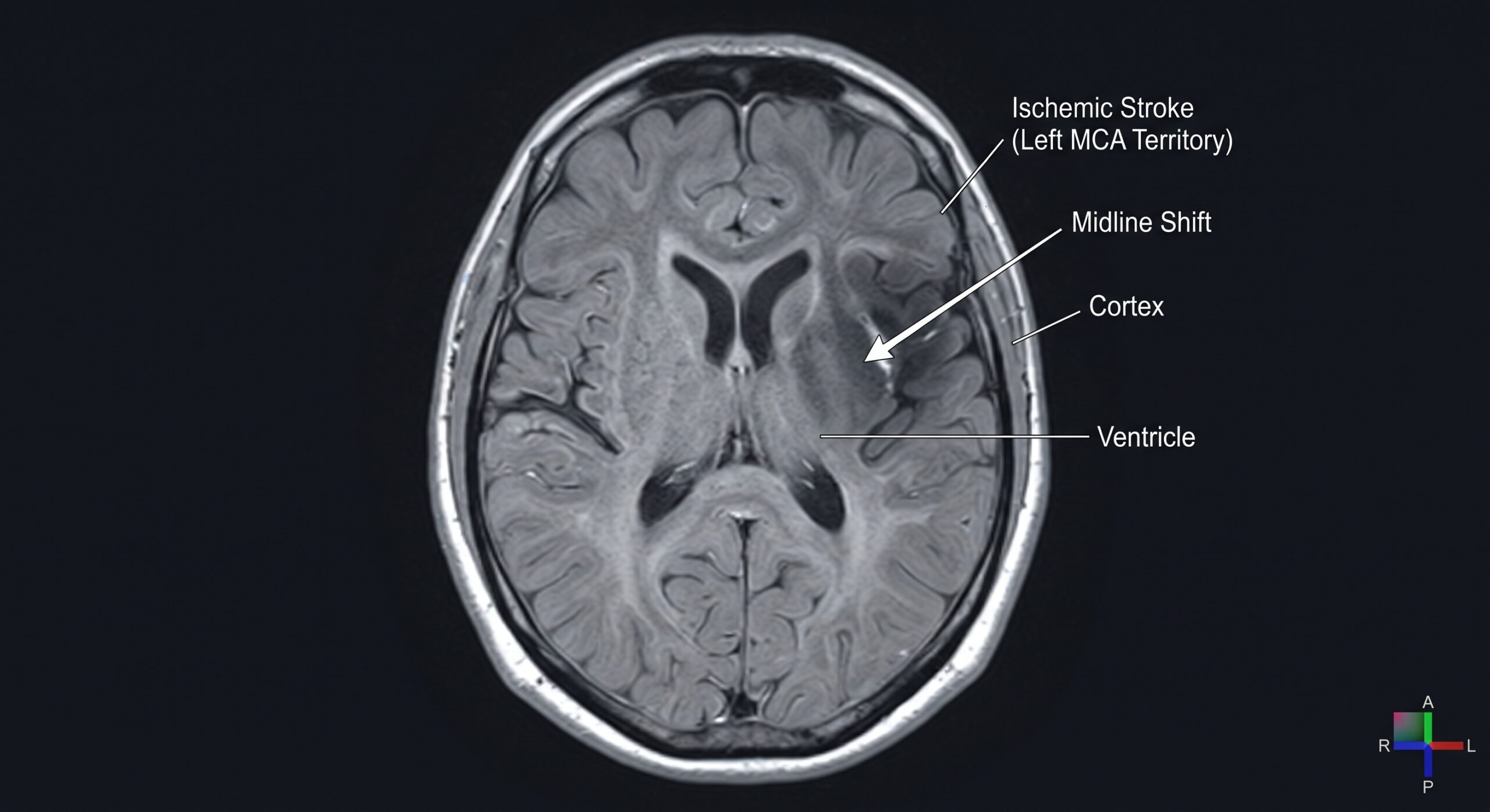
MRI brain scan showing an acute ischaemic stroke. MRI (particularly diffusion-weighted imaging) is far more sensitive than CT for detecting early ischaemic stroke, especially in the posterior circulation — but CT remains the first-line emergency scan because it’s faster and reliably excludes haemorrhage.
CT vs MRI — Which Scan and Why?
Students often get confused about why we use CT when MRI is “better.” Here’s the simple answer: CT is fast, widely available, and brilliant at seeing blood. In an emergency, the first question is “is there a bleed?” — because if there is, thrombolysis is forbidden. CT answers that in minutes. MRI is much more sensitive for early ischaemic change and for posterior circulation strokes, but it’s slower, less available, and the patient has to lie still in a noisy scanner — often impractical for a confused, dysphasic, or deteriorating patient. In practice: CT first, always. MRI later, if the diagnosis is unclear or the posterior circulation is suspected.
Part 4: Your Immediate Nursing Actions — Step by Step
When you suspect a stroke, this is exactly what you do. It’s not complicated, but it must be fast and systematic. Work through it in order — and remember, as a student you must do all of this with a registered nurse. But you can start the clock, take the glucose, and get help.
Immediate Action Checklist
- Note the time. When did symptoms start? If you found them like that, when were they last seen well? Write it down. This single number determines treatment eligibility.
- Call for help. Tell the nurse in charge and the doctor immediately. If the patient is deteriorating — reduced GCS, airway compromise, seizures — put out the 2222 emergency call. Do not wait.
- ABCDE. Work through ABCDE. Airway: are they protecting it? Breathing: give oxygen only if saturations are below 94% — target 94–98%. High-flow oxygen in a non-hypoxic stroke patient can actually worsen outcomes via vasoconstriction. Circulation: check BP, heart rate, rhythm (AF is a major cause).
- Check capillary blood glucose immediately. Hypoglycaemia is the great stroke mimic — and it’s reversible in 30 seconds with glucose. No stroke workup is complete without a glucose reading. If it’s low, treat it — and reassess; the “stroke” may resolve.
- Full set of observations & GCS. Record NEWS2, temperature (fever worsens stroke outcomes — treat aggressively with paracetamol), and a proper neurological set including GCS, pupil reactions, and limb power. Our GCS & neuro observations guide walks you through this.
- Nil by mouth. Do not give food, drink, or oral medication until a trained swallow screen has been done. Aspiration pneumonia is one of the commonest and most dangerous post-stroke complications. If they need oral meds, discuss with the doctor — alternative routes may be needed.
- IV access & bloods. Two large-bore cannulas if possible. Bloods: FBC, U&E, glucose, coagulation screen (essential before thrombolysis), lipid profile, group and save. 12-lead ECG to look for atrial fibrillation or acute cardiac events.
- Positioning. Follow your trust’s policy — this varies. Many hyperacute units keep ischaemic stroke patients lying flat initially to maximise cerebral perfusion, while others elevate the head 15–30°. If there’s any suspicion of haemorrhage or raised ICP, head-up is usual. The key: ask, and follow local policy. Don’t guess.
- Blood pressure — do not lower it yourself. High BP is common after stroke and is often the brain’s way of maintaining perfusion. Acute BP lowering is dangerous unless there’s a specific reason (haemorrhage, thrombolysis candidate with very high BP, aortic dissection). It will be managed by the stroke team. Never give PRN antihypertensives without explicit instruction.
- Document everything. Times, symptoms, obs, glucose, who you called, when they arrived, what was done. This is both good clinical care and your professional protection — see our defensive documentation guide.
Part 5: How to Escalate — The SBAR Script
When you call the doctor or put out the emergency call, use SBAR — Situation, Background, Assessment, Recommendation. It’s the structure that gets you taken seriously and ensures nothing is missed. If you’re nervous about calling, here’s the exact script. Practice it. It works.
S — Situation: “This is [Name], student nurse on [Ward]. I’m with Mr/Mrs [Surname], and I’m concerned they’re having a stroke. Symptoms started at [Time] — that’s [X] minutes ago.”
B — Background: “They were admitted with [reason]. Past medical history includes [AF / hypertension / diabetes if relevant]. They were last seen well at [Time].”
A — Assessment: “On BE-FAST: right-sided facial droop, right arm weakness 3/5, slurred speech. No balance or visual issues noted. GCS 14 (confused), blood glucose 6.2, BP 185/100, pulse 92 irregularly irregular, sats 96% on air, temp 36.8. NEWS2 is [score]. I’ve kept them nil by mouth and put oxygen on standby.”
R — Recommendation: “I need you to review them immediately for the hyperacute stroke pathway, and I think we need to call the stroke team / arrange urgent CT. Can you come now?”
Notice what’s in there: the time of onset first, because that’s what determines everything. A clear BE-FAST breakdown. A glucose result (because hypoglycaemia is the mimic you must exclude). And a clear request. If you only remember one thing: lead with the time of onset. It’s the piece of information the stroke team needs more than any other.
If you’re on shift and need this fast: our Panic Zone has emergency cheat sheets for exactly this scenario — ABCDE, SBAR, and deterioration escalation in one place.
Part 6: Stroke Mimics — When It’s NOT a Stroke
Here’s the mark of a good clinician: you suspect stroke, escalate appropriately — and you also keep the differential in mind. Around 20–30% of suspected “strokes” turn out to be something else. That’s not a failure — it’s better to over-call and be wrong than to miss a real one. But knowing the common mimics makes you a sharper, safer nurse.
| Mimic | How It Looks Like a Stroke | How to Tell |
|---|---|---|
| Sepsis / Infection | Confusion, new falls, “off legs,” drowsiness — especially in the elderly | Check temperature, NEWS2, urine, chest. Sepsis can also co-exist with stroke — don’t assume it’s one or the other. Sepsis red flags guide |
| Hypoglycaemia | Hemiparesis, confusion, reduced consciousness — identical to stroke | Capillary blood glucose — always, immediately. Treat low glucose and reassess |
| Migraine with Aura | Gradual spreading weakness, speech disturbance, visual changes | History of migraine, gradual spread over minutes, younger patient, headache — but never assume; first episode needs imaging |
| Todd’s Paresis | Temporary weakness after a seizure — can last hours | Did they have a seizure? Tongue biting? Incontinence? Witnessed jerking? Resolves over time |
| Bell’s Palsy | Facial droop | Affects the whole side of the face including the forehead. Stroke weakness spares the forehead |
| Functional / Psychological | Weakness, numbness, speech symptoms | Inconsistent examination, Hoover’s sign, give-way weakness — but this is a diagnosis of exclusion; never assume on a ward |
⚠️ The Golden Rule of Mimics
Always assume it’s a stroke until proven otherwise — and “proven otherwise” means a scan and a senior doctor, not your clinical hunch. Even if it turns out to be sepsis or hypoglycaemia, you did the right thing by escalating fast. The only true mistake is the stroke you didn’t call.
Part 7: After the Acute Phase — Complications to Watch For
Most students see stroke patients on the ward after the hyperacute phase — and this is where your observational skills still matter. The first 72 hours are when complications set in, and catching them early changes outcomes. Here’s what to watch:
- Aspiration pneumonia: The commonest serious complication. Every stroke patient must be swallow-screened before any oral intake. If you see a wet, gurgly voice after drinking, coughing on fluids, or a rising temperature and sats drop — suspect aspiration. Keep nil by mouth and escalate.
- Neurological deterioration: A patient whose GCS drops, whose weakness worsens, or who develops a severe headache and vomiting may be having haemorrhagic transformation, brain swelling (oedema), or a second stroke. Repeat neuro obs — any drop in GCS by 2 points is a red flag. See our neurological observations guide.
- VTE — DVT & PE: Immobility after stroke puts patients at high venous thromboembolism risk. Watch for swollen calves, sudden breathlessness, chest pain. Most will be on prophylactic LMWH — check it’s been prescribed and given. Our upcoming VTE guide covers this in depth.
- Pressure ulcers: A patient with reduced mobility and reduced sensation will develop pressure damage fast — within hours, not days. Regular repositioning, pressure-relieving mattress, skin checks. Pressure ulcer guide.
- UTIs & catheter infections: Avoid catheters unless essential. If one’s in, it should come out as soon as possible. A new confusion in a stroke patient is infection until proven otherwise — usually a UTI or chest.
- Shoulder subluxation & pain: A weak, flaccid arm hangs out of the shoulder joint. Correct positioning, supportive slings, and careful handling prevent painful frozen shoulders that handicap rehabilitation.
- Mood & cognition: Around 50% of stroke survivors develop depression or anxiety, and many have cognitive changes — memory, attention, emotional lability (pseudobulbar affect — crying or laughing uncontrollably). These aren’t “just part of it” — they’re treatable. Flag them to the team. Our Wellbeing Hub covers the emotional side — and it applies to patients too.
Part 8: Documentation — What You Must Record
Stroke care is audited relentlessly — the Sentinel Stroke National Audit Programme (SSNAP) reviews every stroke admission, and the timestamps matter. Your documentation needs to be precise and contemporaneous. Record:
- Exact time of onset — or “last seen well at [Time]” if onset unknown
- Exact symptoms — which side, what BE-FAST showed, baseline vs current
- Capillary blood glucose — with time
- Full observations — BP, pulse, rhythm, sats, temp, respiratory rate, GCS, NEWS2
- Who you called, when, and when they arrived
- Actions taken — oxygen, IV access, bloods, ECG, NBM, positioning
- Any changes — improvement or deterioration, with times
As always — if it isn’t documented, it didn’t happen. And in stroke, “didn’t happen” can mean a missed thrombolysis window and a permanently disabled patient. Document as you go, not at the end of the shift. Our defensive documentation guide covers this in full.
Final Thought
Stroke is the emergency where the student nurse at the bedside has more power than almost any other — because you’re the one who sees the patient first, who notices the subtle droop, the slightly slurred word, the unsteadiness on their feet. The consultants and the scanners and the thrombolysis drugs are all useless until someone like you presses the buzzer.
So trust your instincts. If something feels off — if the patient isn’t quite right, if their speech is a bit slower than yesterday, if they can’t stand like they could this morning — do a BE-FAST. Check a glucose. Call for help. You will over-call sometimes. That’s fine. That’s good. The thing you must never, ever do is under-call — because the cost of missing a stroke is measured in millions of dead neurons, and in a person’s independence, and in the life they might have had.
Time is brain. Be the person who starts the clock — and stops the damage.
📚 References & Guidance
- NICE (2022, updated) NG128: Stroke and Transient Ischaemic Attack in Over 16s: Diagnosis and Initial Management. National Institute for Health and Care Excellence — imaging within 1 hour, thrombolysis & thrombectomy windows, swallow screening.
- Intercollegiate Stroke Working Party (2016, reviewed) National Clinical Guidelines for Stroke. Royal College of Physicians — comprehensive UK stroke care standards.
- Sentinel Stroke National Audit Programme (SSNAP) Royal College of Physicians / London School of Hygiene & Tropical Medicine. National audit of hyperacute stroke care and outcomes.
- NHS England (2023) Hyperacute Stroke Services: Service Specification. NHS England / NHS Improvement — HASU network design and time targets.
- The Stroke Association FAST & BE-FAST Public Awareness Resources; UK Stroke Incidence Data. ~100,000 strokes/year, one every 5 minutes.
- Saver JL et al. (2016) “Time is brain: quantified.” Stroke — 1.9 million neurons lost per minute in untreated large-vessel occlusion.
- NICE TA299 / TA431 Alteplase & Tenecteplase for Treating Acute Ischaemic Stroke. Thrombolysis within 4.5 hours of onset.
- MR CLEAN / ESCAPE / EXTEND-IA Trials (2015 onwards) Mechanical Thrombectomy for Large Vessel Occlusion. Evidence base for up to 24-hour thrombectomy windows.
- Purroy F et al. / Nor AMI et al. BE-FAST validation studies — improved detection of posterior circulation strokes vs FAST alone.
- Resuscitation Council UK (2021) Acute Care Standards — Oxygen Therapy. Target 94–98% saturation; avoid unnecessary high-flow oxygen.
- National Institute for Health Research (2020) TIA Early Stroke Risk — ~10% within 7 days, 50% within 48 hours. Urgent TIA review evidence.
- Royal College of Physicians (2017) National Confidential Enquiry into Patient Outcome and Death (NCEPOD): Stroke. Recognition and escalation failures in ward settings.
Written by: Daniel Hancock, Registered Nurse
Focus: Stroke recognition, FAST/BE-FAST, hyperacute pathway, thrombolysis/thrombectomy, nursing management, mimics & complications
Published: October 2026 | Last reviewed: October 2026
Intended for: UK student nurses & newly qualified nurses — ward-based recognition and escalation
Always follow your trust’s specific hyperacute stroke pathway and local policy — positioning, blood pressure targets, and thrombolysis eligibility can vary. This guide is educational and does not replace clinical judgement, senior review, or your trust’s protocols. If you suspect a stroke, escalate immediately — do not delay to read guidance. Time of onset (or last seen well) is the single most important piece of information; record it precisely.
Test Yourself & Build Your Clinical Skills
Stroke recognition is one of the highest-stakes skills you’ll learn. Reinforce it with practice and the frameworks that keep you safe under pressure.